Hip Impingement and Open Surgery
Open Hip Preservation Surgery
Joint Preservation Institute is one of the only centers in Northern California with broad experience in all aspects of hip preservation surgery. Hip preservation is an outstanding option for treating avascular necrosis of the femoral head, cartilage injuries and even localized arthritis.
Surgical Hip Dislocation
One of the great challenges in the treatment of disease of the hip has been limited access to the joint. Due to the multiple powerful muscles that cross the hip and the depth of the joint, dislocation of the hip is usually the only way to access the inner portion of the joint. However, dislocation of the hip can be quite hazardous due to the tenuous blood supply of the femoral head through an artery known as the medial femoral circumflex artery (MFCA). Damage to this blood supply is a major risk factor in the development of avascular necrosis. Professor Reinhold Ganz developed a procedure for safe access to the hip through studies of the anatomy of the MFCA. This artery has been shown to travel up the back part of the neck of the femur and enter the bone at the junction of the head and neck of the femur. Surgical hip dislocation allows access to the joint by using this anatomical information and entering through the front of the joint. In this procedure, an osteotomy (surgical bone cut) is performed on the outer aspect of the femur allowing the joint to be dislocated relatively easily. This procedure provides unlimited access to all aspects of the hip and has made the treatment of cartilage damage to the hip a real possibility. Currently, the procedure has been applied to the treatment of slipped capital femoral epiphysis (SCFE), hip impingement, avascular necrosis, and severe cartilage injuries of the hip.
This procedure provides significant advantage to open procedures of the hip due to the relatively expedient recovery and the substantially decreased blood loss and muscle dissection.
Potential Complications
Surgical hip dislocation can be associated with risk of infection, nerve injury, risk of nonunion or lack of healing at the site of the trochanteric osteotomy, excessive bone formation in the soft tissues, risk of progression of arthritis, and persistent pain. The overall complication rate for major complications is approximately 5%.
CLINICAL CASE : SURGICAL HIP DISLOCATION
The following case is an illustration of the steps involved in surgical hip dislocation in the treatment of CAM impingement.
The patient is a 25 year old athlete with pain during bending activities at his hip. The pain is reproduced by bending the hip up and turning his knee inward (internal rotation of the hip).
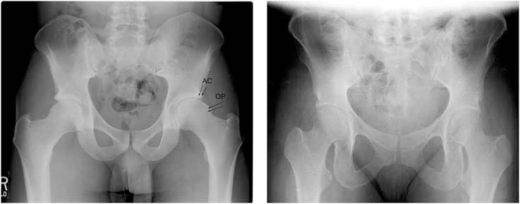
1.These are two radiographs showing the patient's pelvis (left) compared to an a normal pelvis (right). His radiograph reveals a mushroom shaped femoral head with an abnormal contour or osteophyte (OP) which is a classic finding in CAM impingement. The edge of the hip socket (the acetabulum) also has some early arthritis shown by increased bone density at the upper edge (AC).

2.This is the radiograph after the patient has undergone bilateral surgical dislocations. The abnormal bone was resected on the edge of the femoral head. The edge of the acetabulum was also trimmed back to eliminate the abnormal contact.
References
- Philippon MJ, Stubbs AJ, Schenker ML, Maxwell RB, Ganz R, Leunig M. Arthroscopic management of femoroacetabular impingement: osteoplasty technique and literature review. Am J Sports Med. Sep 2007;35(9):1571-1580.
- Tannast M, Kubiak-Langer M, Langlotz F, Puls M, Murphy SB, Siebenrock KA. Noninvasive three-dimensional assessment of femoroacetabular impingement. J Orthop Res. Jan 2007;25(1):122-131.
- Kalberer F, Sierra RJ, Madan SS, Ganz R, Leunig M. Ischial spine projection into the pelvis : a new sign for acetabular retroversion. Clin Orthop Relat Res. Mar 2008;466(3):677-683.
- Larson CM, Giveans MR. Arthroscopic management of femoroacetabular impingement: early outcomes measures. Arthroscopy. May 2008;24(5):540-546.
- Panzer S, Augat P, Esch U. CT assessment of herniation pits: prevalence, characteristics, and potential association with morphological predictors of femoroacetabular impingement. Eur Radiol. Sep 2008;18(9):1869-1875.
- Rakhra KS, Sheikh AM, Allen D, Beaulé PE. Comparison of MRI Alpha Angle Measurement Planes in Femoroacetabular Impingement. Clinical Orthopaedics and Related Research. 2008;467(3):660-665.
- Laude F, Sariali E, Nogier A. Femoroacetabular Impingement Treatment Using Arthroscopy and Anterior Approach. Clin Orthop Relat Res 2009;467:747-752.
- Clohisy JC, St John LC, Schutz AL. Surgical treatment of femoroacetabular impingement: a systematic review of the literature. Clin Orthop Relat Res. Feb 2010;468(2):555-564.
- Barton C, Salineros MJ, Rakhra KS, Beaule PE. Validity of the Alpha Angle Measurement on Plain Radiographs in the Evaluation of Cam-type Femoroacetabular Impingement. Clin Orthop Relat Res. 2011;469:464-469.
- Lukas P Zebala MD, Perry L Schoenecker, M.D., and John C Clohisy, M.D. Anterior Femoroacetabular Impingement: A Diverse Disease with Evolving Treatment Options. Iowa Orthop J. 2007;27:71=81.