Knee Cartilage Repair
Articular Cartilage Injuries in the Knee
Articular Cartilage Injuries
Cartilage is a white, glistening tissue which exists in a number of locations in the body including the ear, nose, ribs, and within the joints. In the joints, it functions to minimize the friction of the bones as they move relative to one another. It is composed predominantly of water but also includes a population of cells and proteins such as collagen. The most important quality of cartilage is its negative electrical charge. This electrical charge attracts water molecules which are responsible for the very good ability of the tissue to lubricate the joint during motion. When the cartilage is damaged, it has a very low healing potential. In some cases, the impact from an injury can lead to complete loss of the cartilage surface from the end of the bones. This is similar in concept to a crater. As one can imagine, the loss of this cartilage “cushion” can lead to much higher pressure on the surrounding areas of cartilage and also lead to damage to other parts of the joint. Such “craters” in the cartilage, particularly when they are large, can lead to significant pain. The source of the pain is unclear as the cartilage itself has no nerve endings. Some experts argue that the “irritable” joint proliferates chemicals which lead to an inflammatory response, This inflammatory response can lead to the classic signs of pain, redness, swelling, and warmth. Others think that the exposed bone is the source of the pain because it is now directly exposed to loads which were dampened previously by the cartilage. A number of treatment are available for cartilage defects. These include microfracture, autologous chondrocyte implantation (ACI), mosaicplasty, and fresh osteochondral allografting.
Arthroscopic Debridement of the Knee
Knee arthroscopy is one of the most commonly performed and most successful orthopaedic surgical procedures. By using small surgical incisions, the amount of soft tissue which is cut is minimized. Additionally, using fiberoptic cameras, even small structures in the knee joint can be easily visualized and treated. Surgical procedures that have been facilitated greatly by knee arthroscopy include debridement or repair of the meniscus, reconstruction of ligaments such as the anterior cruciate ligament (ACL), and treatment of joint (articular) cartilage damage which can result from injuries or arthritis. Typically a number of treatments of the articular cartilage are facilitated with arthroscopy. These include the microfracture procedure, autologous chondrocyte implantation (ACI), osteochondral autografting (mosaicplasty), or fresh osteochondral allografting.
Postoperatively, most patients who have arthroscopy of the knee without additional procedures can return to their activities within three to four weeks.
Microfracture
A technique known as microfracture has demonstrated reliable success in the treatment of articular cartilage damage. This procedure has a number of advantages as well as some limitations. The procedure is aimed at moderately sized lesions of the cartilage up to about 6 square cm which extend down to bone. In the technique, the surgeon debrides the cartilage lesion so that the walls are relatively well defined and vertical in orientation. Then mutiple perforations are made with an angled, pointed tool called an awl. The holes can be approximately 5-8mm apart and 2-3 mm in diameter. As the fluid in the knee is evacuated, the surgeon confirms bleeding into the joint from these holes. The blood from the microfracture has been shown to remodel into a tissue known as fibrocartilage which has similar mechanical properties to normal joint cartilage (also called hyaline cartilage). Currently, fibrocartilage is felt to be inferior in long-term durability to hyaline cartilage but may be adequate for short and medium term pain relief. The effect of the microfracture procedure on the overall progression to global joint arthritis is not known. The great advantage of microfracture is that it can be performed concurrently with an arthroscopy without a second procedure and with no increased muscle dissection. Post-operatively, depending on the specifics of the lesion, the patient is kept on crutches for about six weeks and the use of a continuous passive motion (CPM) is recommended for several hours a day to assist in the nutrition of the newly treated cartilage lesion.
Mosaicplasty
Osteochondral autografting or mosaicplasty is a technique where plugs of cartilage and bone are harvested from non-weight bearing regions of the knee and placed into an area of greater weightbearing. In the case of larger lesions, multiple plugs can be placed next to one another, appearing similar to a mosaic. Similarly to many other techniques, it has a number of advantages and disadvantages. The greatest benefit is that it can be done at the time of the initial procedure and can sometimes be performed using an all arthroscopic technique, facilitating recovery. Additionally, since the tissue is the patient’s own, the risk of disease transmission is eliminated, the graft heals to the host quickly and with no risk of immune-mediated rejection. The disadvantages include a risk of donor site morbidity (pain at the site of graft harvest), the relatively small lesion size that can be treated with each plug, and challenges in matching the radius of curvature of the plug to the area of insertion. In cases where multiple plugs are used, the surface can be quite irregular, leading to increased point pressures in the joint. Additionally, the gaps between the plugs are filled with fibrocartilage which has inferior mechanical properties to normal (hyaline) cartilage.
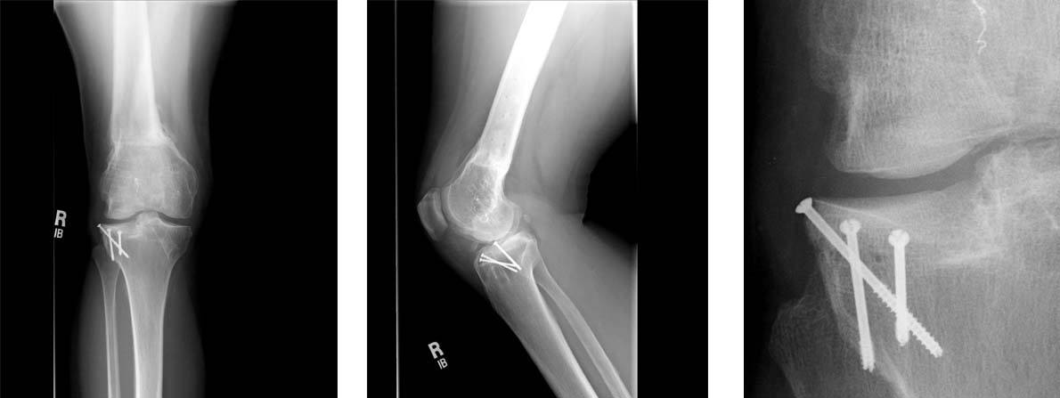
Mosaicplasty Clinical Case
The patient is a 40 year old male with a work related injury to his knee when lifting a 90 lbs suitcase. He had complaints of pain on the inside (medial aspect) of his knee.

1.The standard radiographs of his knee shows an irregularity of the medial femoral condyle (black arrows).

2.This sagittal MRI image shows irregularity of the articular cartilage surfaces in the medial femoral condyle and medial tibial plateau (small white arrows). There is also bone edema (black arrow) noted in the femoral condyle consistent with early degenerative changes. The posterior horn of the medial meniscus appears normal (white arrowhead).

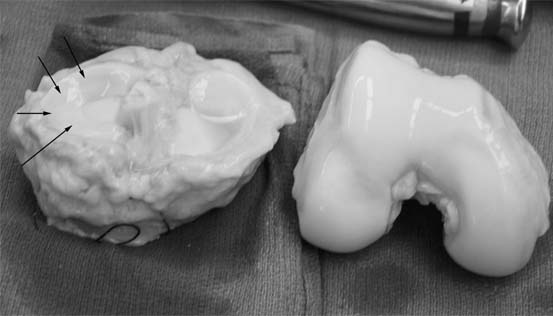
3.The arthroscopic views confirm a full thickness cartilage defect in the medial femoral condyle. The lesion is measured with a cut plastic ruler on the image on the right.

4.Due to the location of the lesion, an open mosaicplasty with one plug is selected as our treatment choice. The harvesting tool is placed within the lesion removing the plug with the damaged cartilage. On the image on the right, the inside of the recipient site is inspected with the arthroscope.

5.In the image to the left, the donor plug (left plug) and recipient plug (right plug) are placed next to each other demonstrating the degree of damage in the recipient site. The image on the right shows the donor plug after being placed flush with the recipient cartilage into the recipient site.

6.On the postoperative xray, the graft can be seen in the medial femoral condyle (black arrow).
Fresh Osteochondral Allografting (Cartilage Transplantation)
Cartilage is the ideal tissue for transplantation as it is protected from the body’s immune system by its fibrous covering. Using a shell of bone as a transportation vehicle, transplantation of cartilage and bone has been performed for over 20 years in the United States. The bone and cartilage composite tissue is termed an osteochondral allograft. The cartilage cells are only able to survive if the tissue is implanted as a fresh graft meaning it has not been frozen or irradiated. These treatments lead to death of the cartilage cells and ultimately failure of the grafts. Prior to 1998, fresh osteochondral allografts were available only through institutional tissue banks. The two largest series of fresh osteochonral allografts in North America are in Toronto and in San Diego.
In San Diego, the procedure has been performed since the early 1980’s. Procedures have been performed predominately in the knee and ankle. The current candidates for the procedure, termed “fresh osteochondral allografting” are those joints where the cartilage damage is so extensive as to not be amenable to the microfracture procedure, autologous chondrocyte implantation, or to the mosaicplasty procedure. Fresh osteochondral allografting is viewed as an intermediate step between these types of procedures and joint replacement surgery. It is suited to patients between age 20 and 40 with relatively major localized damage to the cartilage but an otherwise healthy joint.
The procedure is performed through a small incision measuring between 2 to 3 inches. Fixation of the grafts can be augmented with screws or pins in some cases. After surgery, the patients are on crutches for 6 to 12 weeks. Physical therapy is started immediately after the surgery, focusing on range of motion. We allow weightbearing at the 3 month mark and full activity by 6 months. The cell viability of the cartilage decreases markedly in fresh osteochondral allografts beyond 30 days after graft recovery, making the procedure relatively urgent once a graft becomes available.
The major current challenge to the use of fresh osteochondral allografts relates to the availability of appropriate tissue and the risk of disease transmission. The American Association of Tissue Banks and the FDA under CFR 1270 have established guidelines on donor selection, graft recovery, and processing. Utilizing specific questionnaires, inappropriate donors can be eliminated in 90% of cases. Further, the grafts are tested for a wide spectrum of infective organisms including hepatitis B, C, and HIV as well as bacterial infection which is the most common infecting organism. In 2001, there were a number of deaths associated with allograft transplantations. After further investigation, these have been linked to poor tissue recovery practices and lack of compliance with industry standards. However, in spite of all precautions, the major drawback of fresh osteochondral allografts is the risk of infection.
CASE 1: FRESH OSTEOCHONDRAL ALLOGRAFTING
The following case is an illustration of the steps involved in fresh osteochondral allografting. The patient is a 20 year old male involved in a severe accident two years ago. He has had painful grinding on the outer aspect of his knee and is not able to support his weight on the leg without crutches. He had a previous arthroscopy showing a large cartilage crater on the end of the femur.

1. The arthroscopic photograph above shows a large cartilage defect in the end of the femur on the lateral (outer) side. The size of the defect measured 3.0 x 5.5 cm and involved the underlying bone.

2.MRI images of the knee show a major cavitary defect within the lateral femoral condyle with absence of overlying cartilage.

3.The posterior aspect of the defect is prepared to a precise depth with a specialized cutting device in a cylindrical fashion.

4.A second recipient site is created slightly farther forward on the end of the femur in order to span the entire defect.

5.Both recipient sites (A & B) are treated with precisely contoured grafts covered with healthy cartilage that are removed from a fresh allograft distal femur using specialized instrumentation.
CASE 2: FRESH OSTEOCHONDRAL ALLOGRAFTING
The following case is an illustration of the steps involved in fresh osteochondral allografting. The patient is a 43 year old female with lateral knee arthritis who had previously undergone a distal femoral osteotomy. This was unsuccessful in relieving her pain. Her pain was located on the outer (lateral) side of her knee.

1. These are radiographs of her knee showing minimal arthritis and the presence of the osteotomy plate on the outer aspect of the femur.

2.She initially was evaluated by arthroscopy and found to have extensive cartilage damage on the tibia and on the femur on the outer aspect of the knee. Her, meniscus was almost completely gone.(large arrows). A probe is being used to feel the cartilage of the lateral tibial surface of the knee. The white flaps are evidence of severe damage to the cartilage (small arrow). Based on these findings, she was offered a fresh osteochondral allograft of both the femur and of the tibial surfaces.
3.We obtained a healthy knee graft showing the upper tibia (left) with the meniscus (small arrow) as well as the lower femur (right). The grafts were obtained from a tissue bank. All tissue is obtained from tissue banks with membership in the American Association of Tissue Banks (AATB).
3.We obtained a healthy knee graft showing the upper tibia (left) with the meniscus (small arrow) as well as the lower femur (right). The grafts were obtained from a tissue bank. All tissue is obtained from tissue banks with membership in the American Association of Tissue Banks (AATB).

4.At the time of the allografting procedure, the end of the femur is found to have extensive articular cartilage damage (long thin arrows). The damaged bone and cartilage of the tibia has been removed at this stage of the surgery. (short arrows).

5.At this stage, the tibial graft has been fixed in place with screws (long arrows) along with its meniscus (arrowheads). On the image on the right, a 30 mm diameter femoral condyle allograft plug has been inserted into the area of maximal damage on the femur. (black arrow)

6.Once both grafts are in place, the knee wounds are closed and the meniscus (black arrows) is repaired to the capsule using standard arthroscopic techniques.
7.The final radiographs show the multiple screws used to fix the tibial graft. No fixation was required for the graft on the femur. On the closeup image on the right, one can see the interface between the tibia and the graft (large arrows) as well as the bony surface of the graft on the femur (small arrows). The patient's symptoms of knee pain have nearly fully resolved with the surgery.
CASE 3: FRESH OSTEOCHONDRAL ALLOGRAFTING
The following case is an illustration of the steps involved in fresh osteochondral allografting of the patella. An 18 year old female with previous history of leukemia treated with steroid medications presents with right knee pain for two years. She has difficulty squatting and walking up stairs.

1. These are radiographs of her knee showing cystic areas in both the femur and the tibia (small arrows). These are consistent with a diagnosis of avascular necrosis which can be caused by high dose steroid medications used to treat leukemia.

2.Her MRI images demonstrate severe damage to the surface of the patellar cartilage as well as cystic degeneration within the bone (white arrows)

3.The lesion in the patella is prepared using a guidepin to mark its central portion. Next, a cylindrical recipient site is made and a cylindrical graft is harvested . The graft is then placed in the recipient site and held by a tight fit between the bony surfaces. In the figure to the left, the donor and recipient (with guidepin) patellae are shown. The donor patella has had been cored with a special reamer but the allograft (black arrows) has not yet been removed from the donor patella.

4.These are the followup radiographs at ten months after the surgery. The patient has had substantial improvement in her pain.
FAQ Fresh Osteochondral Allografting
1. What is the success rate of allografting?
The success rate of allografting depends greatly on the quality of the cartilage used and the location of the damaged area. If a high number of the cartilage cells are alive, they will remain within the graft indefinitely.
2. What are the most common complications?
The most common complications are collapse of the allograft or lack of healing at the bone interface between the allograft and the host. Additionally, the rest of the joint can wear out, ultimately, leading to increased pain.
3. Is there rejecation of the grafts?
The osteochondral grafts are composed of both a bony portion (osteo-) and a cartilage portion (-chondral). The cartilage has been shown to elicit minimal immune response in a number of animal studies. The bone contains marrow elements and proteins from the donor which can elicit some immune response. At surgery, we take great efforts to remove as much of the marrow and blood from the graft in order to minimize this response. Once the graft has been in place for 6 months to a year, the bone is gradually remodeled by the recipient and the risk of immune reaction gradually goes down.
4. Do I need to be on immune suppression medications?
Due to the small amount of tissue which generates an immune response (bone), there is no need for immunosuppression after fresh osteochondral allografting.
5. How do they know that the cartilage that is transferred stays alive?
A number of reports have previously showed that the cells remain alive for as long as 14 years. Recently, we revised a patient with a previous fresh osteochondral allograft 29 years ago. He had had a female donor. We analyzed the cells from the region that was grafted and found that indeed there were still female cells within his cartilage.
6. What about disease transmission?
The main risk of disease transmission is bacterial contamination of the grafts. The measures to minimize this risk include the following of strict surgical technique during the recovery of the grafts and efforts to recover the tissue as soon as possible after the donor’s demise. The grafts are cultured to look for bacteria. The risk of viral transmission is also a major concern. The donors are screened based on a standard protocol in order to minimize those with risk factors for blood-borne viral disease. All grafts used at our institution are recovered and processed according to guidelines of the American Association of Tissue Banks (AATB). Further information on the tissue banks that we have used in the past is available at the websites for each of the tissue banks below:
AlloSource
Miami Tissue Bank
Musculoskeletal Transplant Foundation (MTF)
7. Shouldn’t I just have a joint replacement?
The answer to this question depends on your age, expected activity level, the degree of overall cartilage damage, and tissue availability.
8. Isn’t there any way to just inject cells into my joint to heal the cartilage?
No, currently no such technology exists. Currently, promising areas include the use of preseeded matrices, gels, or sponges which can be seeded with the patient’s own cartilage cells or adult stem cells.
9. Why doesn’t the cartilage heal?
Adult cartilage is a highly organized tissue with a paucity of blood vessels which predisposes it to relatively minimal healing response. Adolescent cartilage has much better healing properties that adult cartilage. The healing response of cartilage can be accelerated by stimulating bleeding into the defect using techniques such as the microfracture procedure.
10. How are the grafts kept in place?
The grafts are kept in place using a number of methods. One of these is by a technique known as press-fitting them. With this method, the recipient site is prepared with specialized instruments and a matching set of instruments is used to prepare the donor graft to allow it mechanically lock into the recipient defect. If this fixation is inadequate or not applicable to the area being grafted, the grafts can be held in place using screws or pins.
11. How long will I be in the hospital after fresh osteochondral allografting?
Most patients remain in the hospital for 2 to 3 days after the procedure.
12. When can I start to walk?
You will be maintained on a protective weightbearing regimen for 8 to 12 weeks after the procedure to allow the graft to heal with minimal mechanical stress at the bone-bone interface.
13. What if the allograft fails?
If the allograft fails, the options include repeat allografting if the defect is still relatively small or conversion to some form of prosthetic joint replacement. One of the major advantages of fresh osteochondral allografting is that it does not preclude conversion to a joint replacement at some point in the future.
References
- Steadman JR RW, Singleton SB, et al. Microfracture Procedure for Treatment of Full-thickness Chondral Defects:Technique, Clinical Results and Current Basic Science Status. In Harner CD, Vince KG, Fu FH (eds). Techniques in Knee Surgery. Media, PA, Williams & Wilkins 1999:23–31.
- Bugbee WJ, A.; Chu, C.; and Convery, F. R. Fresh Osteochondral Allografting Of The Patellofemoral Joint. In Proceedings of the 69th Annual Meeting of the American Academy of Orthopaedic Surgeons. Edited, San Francisco, CA, 2001.
- Bugbee WJ, A.; and Rabbani, R. Fresh osteochondral allografting in the treatment of tibiofemoral arthrosis. In Proceedings of the 69th meeting AAOS. Edited, Dallas, Texas, 2002.
- Bugbee W, and Khadavi, B. Fresh osteochondral allografting in the treatment of osteonecrosis of the knee. In AAOS 71st Annual Meeting, pp. #108. Edited, #108, San Fransisco, CA, 2004.
- Jacob R FT, Gautier E, Mainil Varlet P. Autologous osteochondral grafting in the knee: indications results and reflections. Clin Orthop. Clin Orthop;401:170-184.
- Robert H. Chondral repair of the knee joint using mosaicplasty. Orthopaedics & traumatology, surgery & research : OTSR. Jun 2011;97(4):418-429.
- Batty L DS, Bajaj S, Coles B. Autologous chondrocyte implantation: an overview of technique and outcomes. ANZ J Surg. 2011:18-25.
- Falah M NG, Soudry M, et al. Treatment of articular cartilage lesions of the knee. International Orthopedics. 2010;34:621-630.
- Zaslav K CB, Brewster R et al. A prospective study of autologous chondrocyte implantation in patients with failed prior treatment for articular cartilage defect of the knee: results of the Study of the Treatment of Articular Repair (STAR) clinical trial. Am. J. Sports Med. 2009;37:42–55.
- McNickle AG LHD, Yanke AB, Cole BJ. Outcomes of autologous chondrocyte implantation in a diverse patient population. Am. J. Sports Med. 2009;37:1344–1350.
- Gomoll AH, Probst C, Farr J, Cole BJ, Minas T. Use of a type I/III bilayer collagen membrane decreases reoperation rates for symptomatic hypertrophy after autologous chondrocyte implantation. Am J Sports Med. Nov 2009;37 Suppl 1:20S-23S.
- Gikas PD, Bayliss L, Bentley G, Briggs TW. An overview of autologous chondrocyte implantation. J Bone Joint Surg Br. Aug 2009;91(8):997-1006.
- Cole BJ P-GC, Grumet RC. Surgical management of articular cartilage defects in the knee. J. Bone Joint Surg. Am. 2009;91:1778–1790.
- Bekkers J IM, Saris D. Treatment Selection in Articular Cartilage Lesions of the Knee. Am J Sports Med. 2009;37(Suppl. 1):148S-155S.
- Hangody L, Vasarhelyi G, Hangody LR, et al. Autologous osteochondral grafting--technique and long-term results. Injury. Apr 2008;39 Suppl 1:S32-39.
- Steinwachs M KP. Autologous chondrocyte implantation in chondral defects of the knee with a type I/III collagen membrane: a prospective study with a 3-year follow-up. Arthroscopy. 2007;23:381–387.
- Marcacci M KE, Delcogliano M, Filardo G, Busacca M, and Zaffagnini S. Arthroscopic autologous osteochondral grafting for cartilage defects of the knee: prospective study results at a minimum 7-year follow-up. Am J Sports Medicine.